3D Modeling & Printing for Pre-Surgical Planning of Craniopagus Separation
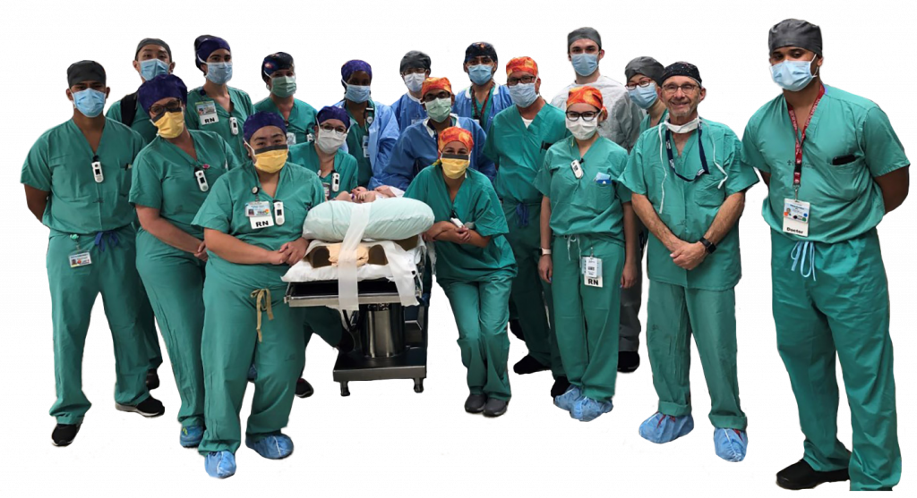
This historic case, which took place October 2020 at UC Davis Medical Center, highlights how a medical illustrator can be integrated into part of the surgical planning process.
Introduction
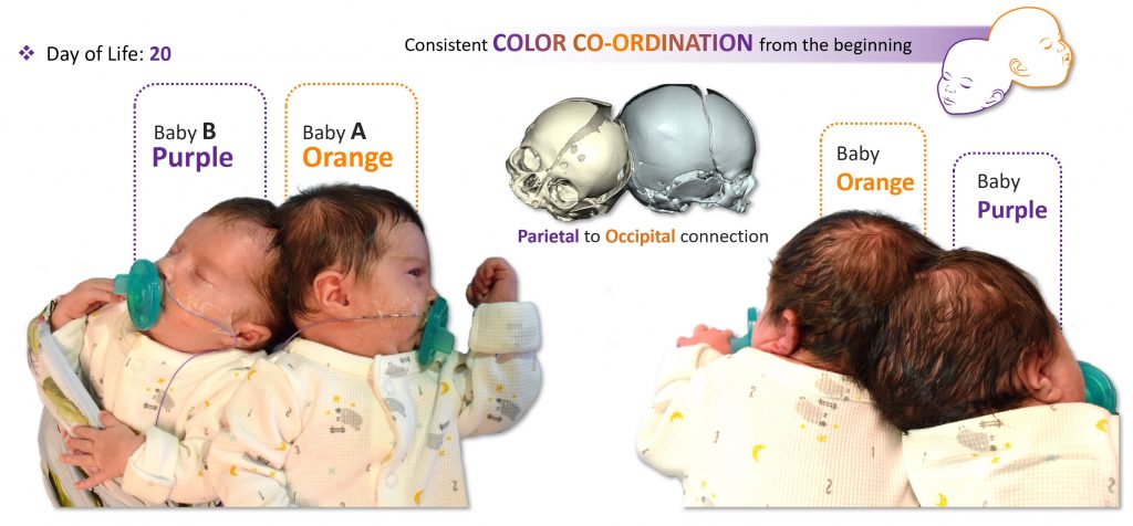
Craniopagus twins, or twins conjoined at the cranium, occur in one out of every ~2.5 million births. Separation, when feasible, requires a multidisciplinary team including neurosurgeons, plastic surgeons, anesthesiologists, and specialized nursing staff. Craniopagus twin girls were born in December 2019. The parietal region of one twin was was joined to the occipital region of her sister. Color coordination was extensively used for operative planning – the twin with the Parietal connection was designated baby Purple, while her sister with the Occipital connection was designated baby Orange.
Methods
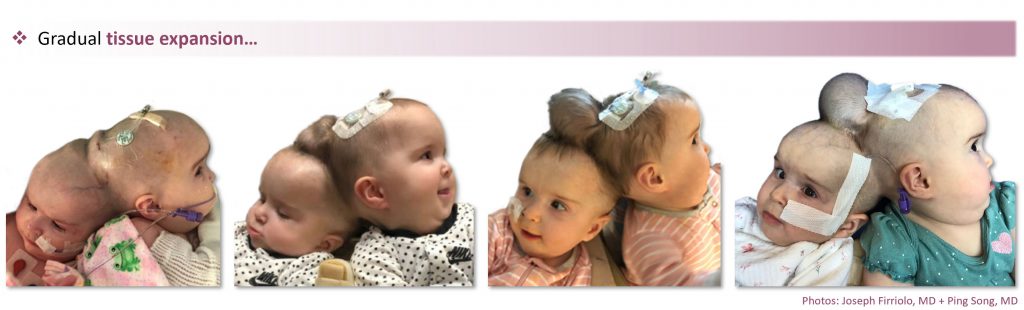
Months prior to the separation, the twins had a custom-designed tissue expander surgically placed to ensure sufficient skin coverage, as separation would otherwise leave each twin with a soft tissue defect. With virtual modeling, the Plastic Surgery team designed a novel wedge-shaped tissue expander with a thin crescentic base, placed at the bony fusion to gain soft tissue for reconstruction.
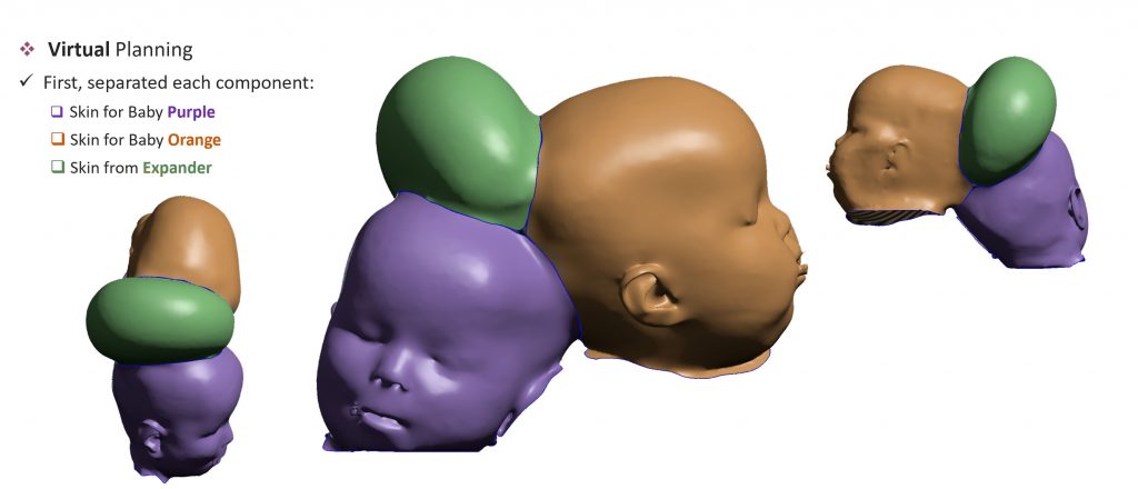
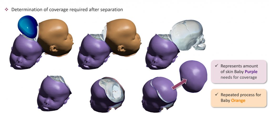
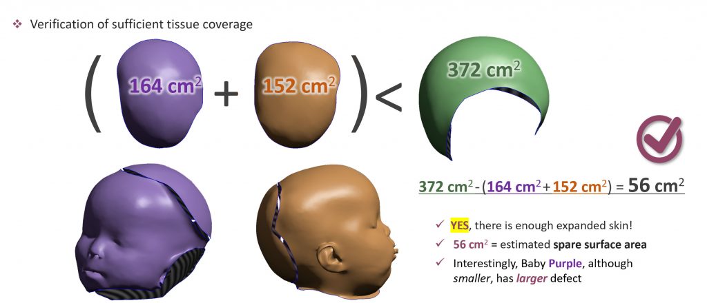
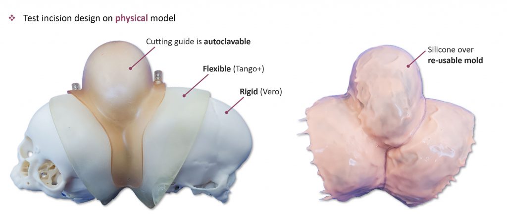
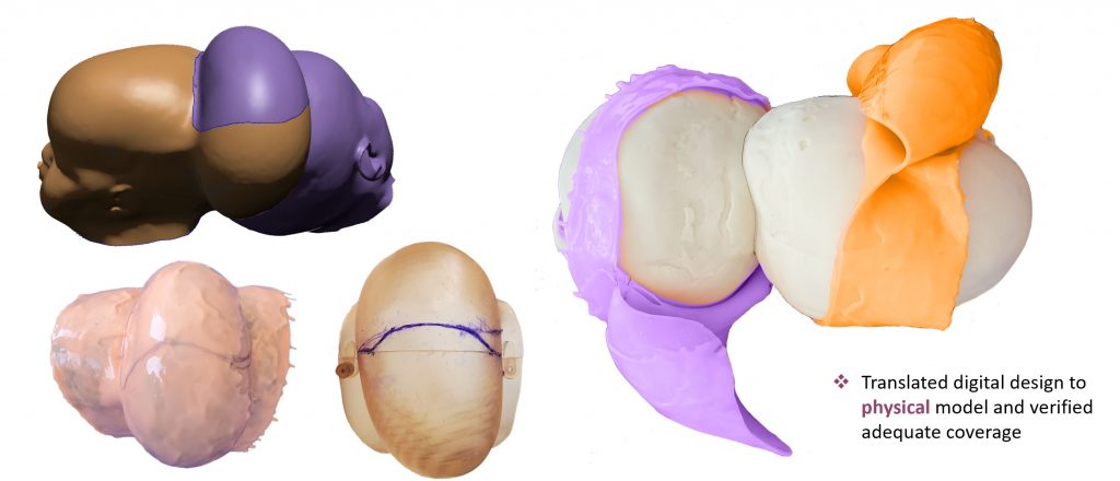
Extensive pre-surgical planning took place prior to the final operation, and included the use of 3D models and prints generated from CT scans. 3D modeling software was used to quantitively verify sufficient skin expansion, as well as facilitate precise skin incision planning. 3D prints were then used to practice the surgery and refine the incision design to ensure optimal skin coverage before ever setting foot in the OR.

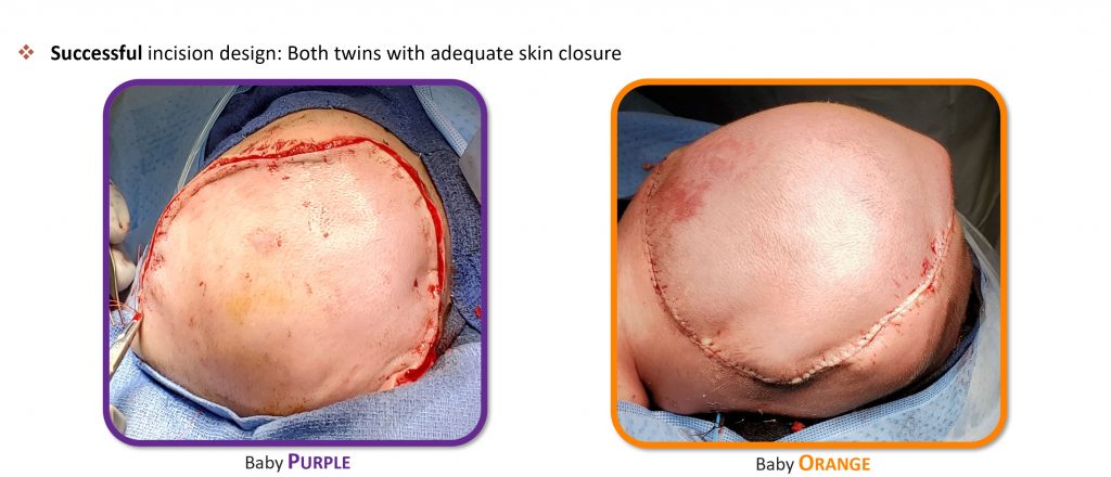
Results
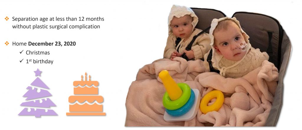
The proposed outline from the models subsequently guided the incision the day of surgery, and ultimately led to a successful surgical outcome. At 10 months of age, the twins underwent cranial separation and successful soft tissue coverage using only native expanded scalp.
Conclusion
Multidisciplinary surgical planning, which can include use of virtual and physical models, is instrumental for rare cases. This type of coordinated team approach can lead to highly successful surgical outcomes.